Testing antibacterial effect of silver nanoparticles on sulfate-reducing bacteria
Summary
In the oil and gas industry, sulfate-reducing bacteria can generate hydrogen sulfide (H2S) in their growth, thus decreasing the commercial value of crude oil, causing metal equipment corrosion and seriously affecting the economic efficiency and the health of workers on drilling rigs. Because of their strong antibacterial and environmental friendly characteristics, silver nanoparticles have been studied and applied in various sectors world-wide. This paper presents some initial research results on the synthesis of silver nanoparticles (NPs) with the mean silver nanoparticle size of 18 - 21nm being effective to reduce bacterial numbers from 106tb/ml to < 10tb/ml (99%). This activity was tested on sulfate-reducing bacteria EPC-KK2 - Desulfomicrobium baculatum which was isolated from crude oil samples taken from Bach Ho field.
Key words: Silver nanoparticle, biocide, sulfate-reducing bacteria, Bach Ho field.
1. Introduction
The sulfate reducing bacteria (SRB) are anaerobic bacteria that produce H2S. This type of bacteria can survive in extreme environments such as high temperature, high pressure, high salinity, to alkaline or acidic environment and is especially quite common in oil fields and exploitation wells. The SRB produce H2S that is acidifying, leading to a reduction in the commercial value of crude oil, corrosion of metalworking equipment, piping, and the health of workers on drilling rigs [1, 2]. Under favourable conditions, desulfurising bacteria can develop biofilm membranes that occlude the reservoir, which reduces the ability of the water to pump.
Currently, popular biocides being used are aldehydes or cyclic amines in combination with cationic active substances, which are highly toxic to humans and the environment. When these biocides are used for a long time the phenomenon of“grease” will occur. It is therefore in need of a new biocide with better antibacterial effect and being more environmentally friendly.
Silver NPs is a new material that is known for its superior disinfection ability and has many applications in life. Silver NPs are very small in size and have very large surface area. They can penetrate easily into microbial cells, alter biochemical mechanisms, inactivate microbial metabolism, and finally destroy them [3].
The antibacterial mechanism of silver NPs is the result of the conversion of silver atoms on the surface of silver NPs to free Ag+ and free ions that will affect bacteria. Some mechanisms of silver NPs action on bacteria are being understood, for example silver ions inhibit the ability of oxygen to travel through the cell wall due to its ability to bind to the peptidoglycan (cell-wall component), leading to cell death [4]. In animals without cell walls, they are unaffected by exposure to these ions. In the other mechanism, silver ions can pass through the cell membrane into the cell and react with sulfhydryl - SH group of the oxygen-converting enzyme molecule, neutralising the enzyme that inhibits bacterial cell respiration. In addition, silver ions are capable of binding to DNA bases and neutralising the charge of phosphate, preventing DNA replication [5 - 8].
Antibacteria with silver NPs is a new research direction in the manufacture of biocides for use in the oil and gas industry. This article presents some results of synthesis of silver NPs and testing bactericidal ability of silver NPs on desulfurising bacteria isolated from Bach Ho crude oil samples.
2. Materials and methods
Materials and equipment
Materials
In this study, all reagents of producing silver NPs colloids were of analytical grades and used as received without further purification. Silver nitrate AgNO3 (99.98%) was used as the silver precursor, which was obtained from Merck (Germany). Sodium borohydride NaBH4 (96.5%) was obtained from Chemical Ltd., whereas the sodium citrate Na3C6H5O7 (99.0%) from Sigma Aldrich was used as producing agents. All the aqueous solutions were prepared in double distilled water.
The improved Postgate’s B bacterial culture medium was used for bacterial culture KH2PO4 0.5g/l; Na2SO4 1g/l; NH4Cl 1g/l; MgSO4 2g/l; NaCl 4g/l; CaCl2 0.1g/l; marine water 200ml; distilled water 800ml; C3H5NaO3 2g/l; pH 7.2g/l; C2H3NaO2.3H2O 1.75g/l;
FeSO4 0.5g/l; yeast extract 0.5g/l; the medium should be autoclaved for 15 minutes at 121oC (250oF). The pH should be adjusted with NaHCO3.
SRB strain was isolated from MSP10 crude oil sample of Bach Ho field and identified by analysing and sequencing 16S rRNA genes.
Equipments
The UV-visible spectra were recorded over the range of 200 - 800nm by Thermo Evolution 300. The size and morphology of silver nanoparticle colloids were investigated using transmitted electron microscope (TEM) Philips CM 120. Particle size measuring device by laser light scattering LA-950.
Methods
The chemical reduction method is used for the production of silver NPs from AgNO3, with the reducing agent NaBH4 or Na3C6H5O7 [9]. The reduction process for silver NPs is shown in Figure 1.
Evaluating the dispersibility of silver NPs: Silver NPs were diluted at different concentrations and UV-VIS spectra were measured to evaluate the existence and contraction of silver NPs.
Isolation and identification of SRB: Bacteria were isolated from Bach Ho oil samples. SRB was isolated on Postgate’s B medium and purified to the mono-assay by means of critical dilution. SRB strain was Gram stained and its cell shape was observed under the optical microscope.
The bacterial strain was cultured and enriched, and identified by analysing and sequencing of 16S rRNA genes.
The biocide efficiency was tested according to bacterial growth after exposure to silver NPs according to API RP38 standard.
3. Results and discussion
Preparation of silver nanoparticle solution
Selection of reducing agents
The results in Figures 2 and 3 show that the particle sizes of products obtained from Na3Cit (Na3C6H5O7) reductant are larger than those from NaBH4. When Na3Cit was used, the absorption wavelength of the silver NPs colloid went up to 450nm under UV-VIS spectrometer and the efficiency of reaction reduced significantly comparing to NaBH4.
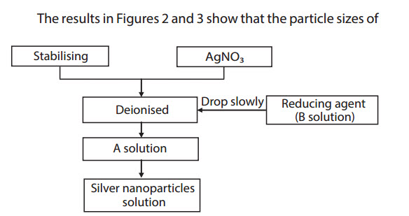 |
Figure 1. Diagram of silver nanoparticle preparation
 |
Figure 2. UV-VIS spectrophotometric and TEM image of silver NPs with Na3Cit
 |
Figure 3. UV-VIS spectrum and TEM image of silver NPs with NaBH4
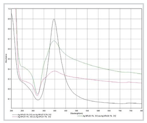 |
Figure 4. UV-VIS spectra of silver nanoparticle samples (AgNPs 30, 31, 33)
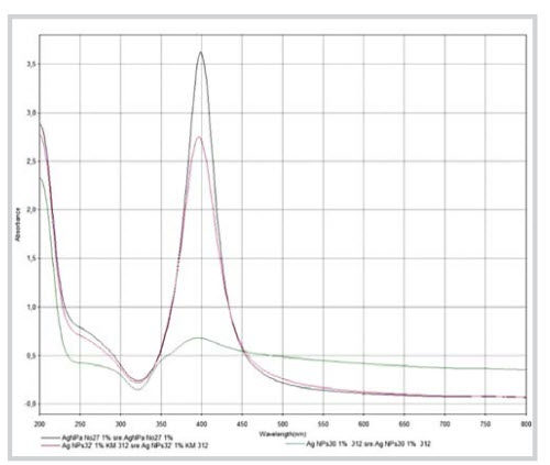 |
Figure 5. UV-VIS spectra of silver NPs solution samples with concentration ratios stabilised/AgNO3: 0.05; 0.275 and 0.5
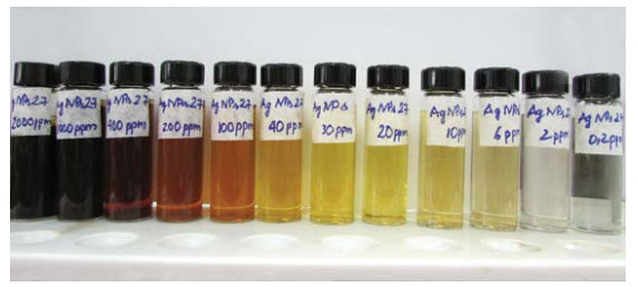 |
Figure 6. Silver NPs solution in different concentrations
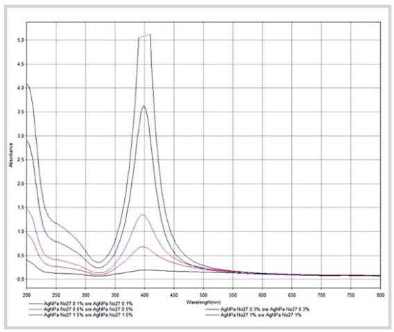 |
Figure 7. UV-VIS spectra of silver nanoparticles AgNPs 27 at concentrations of 0.1 , 0.3 , 0.5 , 1 and 1.5
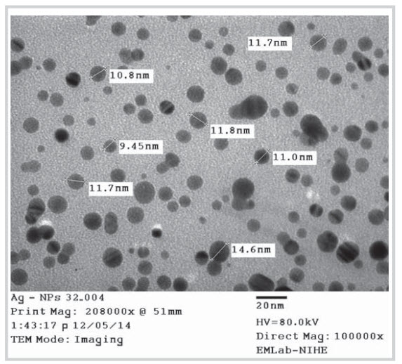 |
Figure 8. TEM of AgNPs 32
It means that the reduction capacity of Ag+ ions to Ago by NaBH is higher than Na Cit. As a result, NaBH was comparing to AgNO3. Figure 5 shows the UV-VIS spectrographic chromatograms of silver NPs colloids with NaBH4 to AgNO3 ratios being 2:1; 3:1; 4:1 at 0.380; 0.900; 0.683 peak heights respectively. It means that the content of reducing agents would considerably affect the numbers of silver NPs produced (Figure 4). The results showed that the highest concentration of silver NPs colloid was chosen for the synthesis of silver NPs.
Silver nanoparticle preparation conditions
- Effect of NaBH4/AgNO3 concentration ratio
In order to achieve the highest reaction efficiency, the NaBH4 reductant must be used in excess amount obtained at NaBH4 to AgNO3 ratio of 3:1.
- Effect of the stabilising agent/AgNO3 ratio
The results in Figure 5 indicated that the UV-VIS absorption peak height of products obtained from a 0.05 ratio of stabilising agent to AgNO3 is much lower than that from 0.275 and 0.5 ratios. It can be explained that too low stabilising agent concentration will not create mixed molecular surrounding silver NPs, and this is the reason why the nanoparticle size increases and the number of silver NPs decreases. The highest UV-VIS absorption peak was obtained from the 0.5 ratio of stabilising agent to AgNO3.
Evaluation of silver NPs colloid characteristics
Silver NPs colloids were prepared at different concentrations from 0.2 to 2,000ppm (Figure 6). UV-VIS absorption spectrometer of range of silver NPs concentrations was shown in Figure 7. The diluted samples all reached a peak of 400nm which indicated that the properties of silver NPs were not affected by the dilution in water.
The TEM image of silver NPs at 2,000ppm (Figure 8) showed that the size of silver NPs was at approximately 15 ± 4nm. These nanoparticles meet the requirements of size which is effective for biocide (< 100nm).
Figure 9 showedtheresultsofsizedistribution of silver NPs at concentration, 2000ppm, and the mean size is around 19nm.
Isolation and identification of bacteria
The EPC-KK2 sulfate reducing strain was isolated from oil sample MSP10-1014. On the microscope, these cells are short rod shaped, Gram-negative bacteria, planktonic. This type of bacteria is anaerobic, and anaerobic respiration uses sulfate as the final electron acceptor, producing H2S. They grow on lactate substrates. Optimum conditions of growth include: appropriate temperature is 30 - 32oC; pH is 7.5; and salt concentration of NaCl is 2.5g/l.
The EPC-KK2 strain was classified and identified by analysing and sequencing 16S rRNA genes.
The results of gene analysis (Figure 10) of EPC-KK2 showed that the amplified genomes were similar to Desulfomicrobium baculatum KF536747.1 (Figure 11). From the results of sequencing 16S rRNA genes and the cell morphology (Figure 12), EPC-KK2 can be identified as Bacteria; Proteobacteria; Deltaproteobacteria; Desulfovibrionales; Desulfomicrobiaceae; Desulfomicrobium baculatum.
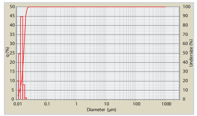 |
Figure 9. Size distribution of AgNPs 27 in solution
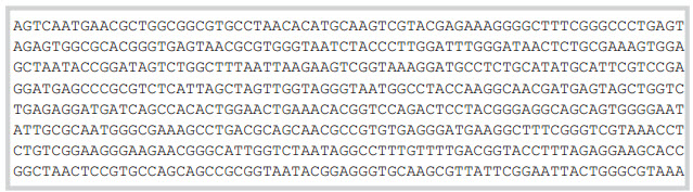 |
Figure 10. Sequence genes of strain EPC-KK2
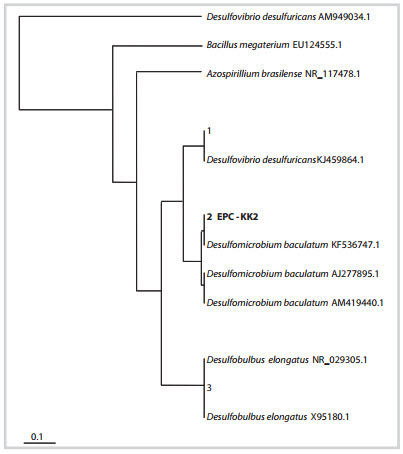 |
Figure 11. Phylogenetic position of EPC-KK2 species with relatives based on16S rRNA sequences General characteristics of the bacterium Desulfomicrobium baculatum:
Cells: rod or ellipsoidal-shaped cells, 0.5 - 0.9 x 1.3 - 2.9μm, with round ends, either singly or in pairs [10]. Gram- negative stain reaction and having cell- wall structure. Cells are motile, usually by a single polar flagellum. Endospores are not formed.
Growth condition: anaerobic, pre- reduced medium or reducing agent required in medium for growth. Growth can occur by anaerobic respiration with sulfate or sulfoxy-anions as terminal electron acceptor, producing H2S.
Optimal temperature from 25 - 30oC.
Substrates used: simple organic compounds that serve as electron donors during sulfate respiration include lactate, pyruvate, ethanol, formate and hydrogen. Sulfate respiration with lactate as electron donors is incomplete, with the formation of acetate and CO2. Hydrogenase is present. Cells contain b- and c-type cytochromes. Metabolism can also be fermentative on simple organic substrates, including pyruvate, malate or fumarate; carbohydrates are not fermented. No specific vitamins are required [10].
Antibacterial effect of silver NPs
There are a number of theories on the antibacterial ability of silver NPs colloid, in which the absorbed theory is most convincing. The main idea of the theory is that the bacterial cell is inactive due to the electron storage between the negative charged surface of the cell and the Ag+ ions which have been absorbed to it. These ions would penetrate into the inside of the bacterial cell and inactivate them.
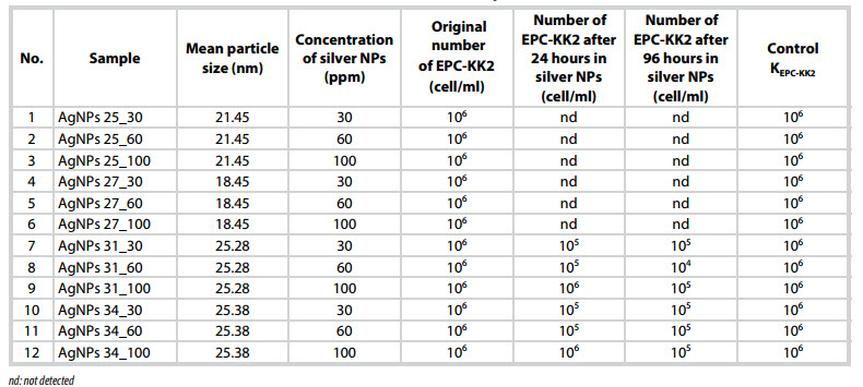 |
Table 1. The effect of NPs size and concentration of AgNPs colloids on EPC-KK2
 |
Table 2. Test on antibacterial concentrations of AgNPs 25 with EPC-KK2
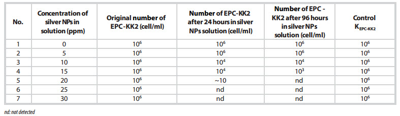 |
Table 3. Test on antibacterial concentrations of AgNPs 27 with EPC-KK2
The silver NPs samples collected are tested with the antibacterial effect based on their particle sizes. According to announced studies, the size of silver NPs is an important factor that affects the antibacterial property. For different size of silver NPs, the activities of the solution on the bacteria are very different [8]. Overall, the size of the silver NPs that is effective for bacteria is within the range of 1 to 50nm. For the isolated strain, the size of silver NPs is within 1 - 50nm. The effective concentration of silver NPs solution on different bacteria varies. Normally, the minimum inhibited concentration of silver NPs solution is about 10 - 75ppm. The silver NPs solutions which are prepared and tested in the study include: AgNPs 25, AgNPs 27, AgNPs 31, and AgNPs 34. These solutions did not show precipitation and were tested with the EPC-KK2 type. The results are shown in Table 1.
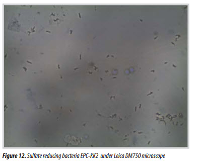 |
Figure 12. Sulfate reducing bacteria EPC-KK2 under Leica DM750 microscope
Table 1 shows the ability of silver NPs colloid AgNPs 25 and AgNPs 27 to kill EPC-KK2 strain with the mean particle size < 25nm. AgNPs 31 and AgNPs 34 with the mean particle size > 25nm did not kill EPC-KK2 strain, effectively.
With the silver NPs size < 25nm, the antibacterial effect of AgNPs 25 and AgNPs 27 were observed at different concentration between 0 - 30ppm. The results in Tables 2 and 3 show that EPC-KK2 was not observed after the experiment with AgNPs 25 and AgNPs 27 samples at the concentration of 20ppm.
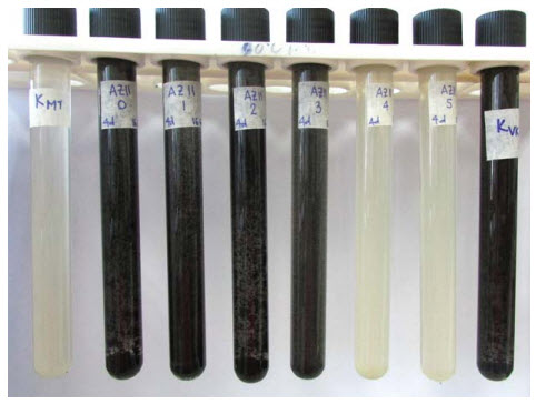 |
Figure 13. EPC-KK2 strain grown again in test of silver NPs
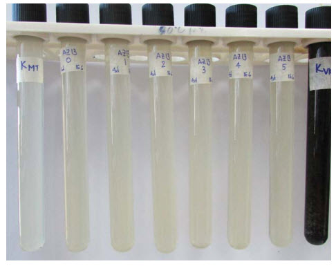 |
Figure 14. EPC-KK2 strain killed in test of silver NPs
Pictures of the cultivated EPC-KK2 strain in the experiment to assess the antibacterial ability of silver NPs colloid are shown in Figures 13 and 14.
The results of the tests show that the antibacterial ability of AgNPs 27 and AgNPs 25 samples are highly effective at the concentration of 20ppm for the EPC- KK2 strain.
4. Conclusion
The EPC-KK2 strain, which was isolated from the Bach Ho crude oil samples and classified as Desulfomicrobium baculatum, was used to evaluate the antibacterial effectiveness of silver NPs colloid. Silver NPs (mean silver NPs size 18 - 21nm) were effective to reduce the antibacterial activity from 106tb/ml to < 10tb/ml (99%) at 20ppm. The results showed that silver NPs are highly effective in killing SRB. This would create a new approach for applying silver NPs as biocide in Vietnam's oil and gas industry. References
1. Lai Thuy Hien, Dang Phuong Nga. Physiological and biochemical properties of some sulfate-reducing bacteria strains isolated from Bach Ho oil field. Journal of Biology. 1998; 20(2): p. 33 - 38.
2. Lai Thuy Hien, Le Phi Nga. Study on metal corrosion potential of Desulfovibrio vulgaris. Journal of Biology. 1992; 14(4): p. 26 - 29.
3. Catalina Marambio-Jones, Eric M.V.Hoek. A review of the antibacterial effects of silver nanomaterials and potential implications for human health and the environment. Journal of Nanoparticle Research. 2010; 12(5): p. 1531 - 1551.
4. T.A.Brown, D.G.Smith. The effects of silver nitrate on the growth and ultrastructure of the yeast Cryptococcus albidus. Microbios Letters. 1976; 3: p. 155 - 162.
5. M.R.Richards, H.A.Odelola, B.Anderson. Effect of silver on whole cells and spheroplasts of a silver resistant Pseudomonas aeruginosa. Microbios. 1984; 39(157 - 158): p. 151 - 157.
6. Yakabe Yoshikuni, Sano Takayuki, Ushio Hidetoshi, Yasunaga Tatsuya. Kinetic studies of the interaction between silver ion and deoxyribonucleic acid. Chemistry Letters. 1980; 9(4): p. 373 - 376.
7. Reed M.Izatt, James J.Christensen, J.Howard Rytting. Sites and thermodynamic quantities associated with proton and metal ion interaction with ribonucleic acid, deoxyribonucleic acid, and their constituent bases, nucleosides, and nucleotides. Chemical Reviews. 1971; 71(5): p. 439 - 481.
8. Gianluigi Franci, Annarita Falanga, Stefania Galdiero, Luciana Palomba, Mahendra Rai, Giancarlo Morelli, Massimiliano Galdiero. Silver nanoparticles as potential antibacterial agents. Molecules. 2015; 20(5): p. 8856 - 8874.
9. Shuai He, Honglin Chen, Zanru Guo, Biqing Wang, Chongli Tang, Yujun Feng. High-concentration silver colloid stabilized by a cationic geminisurfactant. Colloids and Surfaces A: Physicochemical and Engineering Aspects. 2013; 429: p. 98 - 105.
10. George M.Garrity. Bergey’s manual of systematic bacteriology (2nd edition). Springer Science & Business Media, USA. 2001; 2: p. 944 - 945.